Academic Profile

Currently seeking postdoctoral opportunities (2026-2027)

Johan Ajnabi

Mechanotransduction · Epigenetic Regulation · Tissue regeneration

I am Johan Ajnabi, a PhD candidate in the Jamora laboratory at BRIC-inStem, studying how injured skin converts physical cues into signaling, chromatin-state change, and epidermal repair programs.

My work brings together primary keratinocyte systems, fluorescence imaging, molecular biology, spatial transcriptomics, and mechanobiology approaches to understand tissue repair and disease-associated remodeling.

Flagship Work

Representative projects and outputs

The strongest entry points into my current research program are the projects below: mechanotransduction-driven epigenetic regulation, spatial immune-state mapping, and epithelial stemness signaling.

2026 preprint

Mechanotransduction and DNMT3A during cutaneous wound repair

Defined how wound-induced mechanical cues regulate nucleocytoplasmic partitioning of DNMT3A through actin remodeling and ERK1/2 signaling during epidermal repair.

Spatial transcriptomics

Immune-state mapping in vitiligo skin after NB-UVB treatment

Profiled treatment-associated remodeling of the cutaneous immune landscape using spatial transcriptomic analysis complemented by validation in patient tissue.

Stemness and signaling

Mindin-integrin-STAT3 control of keratinocyte stemness

Contributed to work uncovering a signaling axis that sustains keratinocyte stemness through integrin endocytosis and downstream transcriptional activation.

Prefer a narrative view of the work? Browse the flagship project pages.


Research Pillars

Three connected directions

My work is organized around how skin cells sense tissue-scale change, translate those cues into molecular programs, and remodel during repair or disease.

Pillar 01

Mechanical regulation of chromatin

I study how wound-associated mechanics, actin remodeling, and ERK signaling alter the localization and function of chromatin regulators such as DNMT3A.

Pillar 02

Tissue regeneration and epithelial repair

My research focuses on how epidermal cells coordinate migration, repair, and state transitions in wounded tissue and primary keratinocyte systems.

Pillar 03

Inflammation, stemness, and translational biology

I connect regeneration-focused questions with spatial transcriptomics, immune-state remodeling, keratinocyte stemness, and disease-relevant epithelial biology.


Research Alignment

Where I want to take the work next

I am looking for postdoctoral environments where mechanobiology, epigenetic regulation, tissue regeneration, and translational biology can be brought together in ambitious ways.

Questions I want to pursue next

  • How mechanical microenvironments influence chromatin regulator localization and cell fate.
  • How repair programs transition between regeneration, inflammation, and fibrosis.
  • How tissue architecture shapes epithelial plasticity in injury and disease.

Model systems and methods I bring

  • Primary epithelial and keratinocyte culture, wound repair biology, and tissue-section analysis.
  • Live-cell imaging, biochemical assays, genetic perturbation, qPCR, and validation workflows.
  • Spatial transcriptomics and interpretation of tissue-level expression states.

Research environments where I fit well

  • Interdisciplinary groups bridging cell biology, tissue repair, and regenerative medicine.
  • Labs combining mechanobiology with epigenetics, imaging, or systems-level profiling.
  • Collaborative teams with translational interest in regeneration, tissue repair, or disease remodeling.

Technical Toolkit

Methods and systems

A compact view of the experimental and analytical toolkit I bring to postdoctoral work.

Imaging and biophysics

Dynamic readouts of localization, movement, and tissue architecture.

Confocal imaging Live-cell imaging Multiphoton microscopy FRAP FRET Immunohistochemistry Immunofluorescence

Cell and molecular biology

Experimental systems for perturbing signaling, chromatin, and epithelial states.

Primary keratinocytes Mouse models Mechanobiology assays Genetic perturbation Biochemical assays Co-immunoprecipitation (Co-IP) Chromatin immunoprecipitation (ChIP) qPCR validation Western blotting Flow cytometry

Analysis and visualization

Quantitative interpretation, image analysis, and publication-ready data display.

Tissue-section analysis FIJI/ImageJ analysis GraphPad Prism Bioinformatics interpretation Basic R and Python Basic web development (HTML/CSS/JS)

Research Experience

Research experience and training

Training across epidermal repair, stem cell biology, tissue analysis, and plant molecular biology.

Jul 2024 – Present Research Assistant

Shiv Nadar Institute of Eminence

Delhi–NCR, India · Supervisor: Prof. Colin Jamora

Primary focus

Epigenetic and mechanical regulation of the cutaneous wound response, with a focus on wound-induced regulation of DNMT3A.

Model systems

Primary mouse keratinocytes for studying mechanotransduction-driven nuclear localization and function of epigenetic regulators.

Aug 2019 – Present Research Scholar

BRIC–Institute for Stem Cell Science and Regenerative Medicine (inStem)

Bengaluru, India · Supervisor: Prof. Colin Jamora

Primary project

Mechanotransduction-driven regulation of DNMT3A localization during epidermal repair, integrating molecular biology, biochemistry, fluorescence microscopy, and genetic engineering.

bioRxiv (preprint), 2026 [Ajnabi et al., 2026]

Tissue-scale and translational biology

Applied spatial transcriptomics to investigate treatment-responsive molecular programs in vitiligo and explored extracellular matrix regulation of cutaneous fibrosis using transgenic mouse models, integrating tissue-scale profiling with mechanistic validation.

Clinical & Translational Immunology, 2026 [Dutta et al., 2026] · JID, 2023 [Rana et al., 2023]

Stemness and niche regulation

Contributed to the discovery of a Mindin–integrin–STAT3 signaling axis that sustains keratinocyte stemness through integrin endocytosis, and to studies demonstrating how autocrine Mindin signaling maintains stem and progenitor states in skin epithelium and carcinoma cells.

Cell Communication and Signaling, 2026 [Dam et al., 2026] · Cell Reports, 2022 [Badarinath et al., 2022]

Host-pathogen interactions

Investigated LL-37-mediated inhibition of SARS-CoV-2 entry using mammalian cell systems, pseudovirus assays, and flow cytometry.

Frontiers in Immunology, 2023 [Bhatt et al., 2023]
Jan 2018 – Jul 2019 AIEEA-PG Scholar

ICAR–National Institute for Plant Biotechnology

New Delhi, India · Supervisor: Dr. Monika Dalal

Master's research

Identified cis-regulatory regions controlling PM19 gene expression in wheat.

MSc Thesis, ICAR–IARI, 2019 [Ajnabi, 2019]

Methods

In-silico genome analysis, gene/promoter cloning, transient GUS assays, plant tissue culture, and plant transformation.


Awards & Recognition

Awards, fellowships, and qualifications

Competitive awards, fellowship support, and national qualifying examinations that have shaped my academic training.

Awards

  • Paeonia Foundation Travel Award MBI Conference, National University of Singapore · 2023

    Competitive travel award supporting presentation of mechanobiology research at Mechanobiology in Health and Disease, National University of Singapore.

Scientific Community

  • Featured by Epithelial Mechanics Fan Club Research Spotlight · 2026

    Research on actin-dependent mechanotransduction and DNMT3A-mediated epigenetic regulation during cutaneous wound repair was highlighted by the Epithelial Mechanics Fan Club community. Read feature

Fellowships

  • ICMR Junior Research Fellowship Life Sciences · 2019 Indian Council of Medical Research (ICMR), India
  • DBT-BCIL Junior Research Fellowship Biotechnology · 2019 Department of Biotechnology (DBT), Government of India
  • CSIR Junior Research Fellowship Life Sciences · AIR 20 · 2019 Council of Scientific and Industrial Research (CSIR), India
  • ICAR AIEEA-PG Scholarship Plant Biotechnology · AIR 1 · 2017 Indian Council of Agricultural Research (ICAR), India

Qualifications

  • ASRB-NET Agricultural Biotechnology Lectureship · 2020 Agricultural Scientists Recruitment Board (ASRB), India
  • Joint CSIR-UGC NET Life Sciences · AIR 20 · 2019 Council of Scientific and Industrial Research (CSIR) and University Grants Commission (UGC), India
  • GATE XL Life Sciences · AIR 8 · 2019 Graduate Aptitude Test in Engineering, administered nationally by IITs/IISc
  • ICAR AICE-JRF/SRF Agricultural Biotechnology · AIR 3 · 2019 Indian Council of Agricultural Research (ICAR), India

Publications

Selected publications and full record

Selected work below highlights the strongest links to my current research identity: mechanotransduction, tissue regeneration, spatial profiling, and epithelial stemness.

First-author preprint

Preprint

Actin-dependent mechanotransduction controls DNMT3A partitioning during wound healing

Defines a mechanotransduction-driven route from wound-induced actin remodeling and ERK1/2 signaling to nucleocytoplasmic control of an epigenetic regulator.

Spatial transcriptomics

Clinical & Translational Immunology

Spatial transcriptomic analysis of NB-UVB-treated vitiligo skin

Uses spatial transcriptomics and validation in patient tissue to resolve treatment-linked immune-state remodeling in the cutaneous landscape.

Stemness signaling

Cell Communication and Signaling

Mindin-mediated integrin endocytosis activates STAT3 to maintain keratinocyte stemness

Connects extracellular matrix signaling, integrin trafficking, and transcriptional activation to the maintenance of keratinocyte stemness.

Epithelial plasticity

Cell Reports

Snail maintains epithelial stem/progenitor states through Mindin signaling

Identifies an autocrine Snail-Mindin signaling loop that sustains epithelial stemness in normal skin and carcinoma-associated contexts.

Full publication record

Filter by article type or sort chronologically below. A PDF version is also available in CV (PDF).

2026

Dam, B., Ajnabi, J., Saha, T., Shrivastava, A., Badarinathan, K., Hegde, A., Dutta, A., Jasoria, S., Kataria, S., Singh, A., Jamora, C.
Mindin-mediated αM-integrin endocytosis activates STAT3 to maintain keratinocyte stemness
Cell Communication and Signaling, 2026

Abstract

Background: Keratinocyte stem cells are essential for maintaining epidermal homeostasis and enabling efficient tissue repair. Regulation of their self-renewal and differentiation is critical, as its disruption can impair regeneration and drive pathological conditions such as chronic wounds and cancer. We previously identified the matricellular protein Mindin as a key regulator of keratinocyte stemness through its interaction with the αMβ2 (CD11b/CD18) integrin and subsequent activation of the transcription factor STAT3. However, the mechanism connecting Mindin and integrin at the cell surface to the intracellular activation of STAT3 remained undefined.

Methods: We employ biochemical and imaging analysis along with molecular dynamics simulations to dissect Mindin–integrin–STAT3 signalling in primary mouse keratinocytes. Stemness of epidermal keratinocytes are assessed using bulk RNA sequencing, quantitative PCR, and cell-based assays.

Results: Our work demonstrates that the F-Spondin domain of Mindin constitutes the minimal integrin-binding module required to initiate downstream signalling. F-Spondin binding to the integrin at the plasma membrane does not elicit the full activation state of the integrin. Instead, it promotes Src-kinase dependent endocytosis of the integrin receptor to the early endosomes. Analysis of integrin conformational dynamics reveals that the acidic environment of early endosomes is essential to achieve a signalling-competent state. This mechanism extends to pathological contexts, as we demonstrate a requirement for endocytosis in activating STAT3 signalling and preserving stem-like properties in a cancer stem cell model.

Conclusions: These findings highlight a previously unrecognized layer of spatial control in integrin signalling, confirming endosomal trafficking as a critical determinant of stem cell behaviour and offering new conceptual and therapeutic opportunities across regenerative biology and cancer.

My contribution

I performed the confocal imaging and its analysis, and carried out the western blotting for this study.

Techniques

Confocal imaging Western blotting
Ajnabi, J., Dam, B., Gupta, E., Saha, T., Dutta, A., Kumar, S., Gupta, A., Palakodeti, D., Jamora, C. ★ First author
Actin-dependent mechanotransduction controls nucleocytoplasmic partitioning of DNMT3a through ERK1/2 signaling during cutaneous wound healing
bioRxiv (preprint), 2026

Abstract

Cutaneous wound healing is a multifaceted physiological process that requires a cell state transition from homeostasis to tissue repair. An important contributor to this reprogramming is the wound-induced mechanical cues that are perceived by epidermal keratinocytes. Previously we identified that the nuclear translocation of the de novo DNA methyltransferase 3A (DNMT3a) upon wounding as an important regulator of this cell-state transition. However, the molecular mechanisms linking mechanotransduction to epigenetic regulation remain incompletely understood. Here we show that under homeostasis, active ERK1/2 phosphorylates DNMT3a, intramolecularly masking its nuclear localization signal (NLS), resulting in its cytoplasmic sequestration. Upon wounding, actin cytoskeleton remodeling leads to the downregulation of the Extracellular Signal-Related Kinase 1/2 (ERK1/2) pathway. Wound-induced ERK1/2 inactivation unmasks the NLS, enabling DNMT3a nuclear translocation. Collectively, these findings define a mechanotransduction-driven signaling axis linking cytoskeletal dynamics to epigenetic regulation and confirms an active role for differentiated keratinocytes in initiating early wound repair programs.

My contribution

As first author, I led this study and performed all of the experimental work except the molecular dynamics simulations.

Techniques

Immunofluorescence Confocal imaging Western blotting Mechanobiology assays
Dutta, A., Gupta, D., Ajnabi, J., Dam, B., P, S., Jamora, C.
Spatial transcriptomic analysis of the immune landscape following NB-UVB treatment of vitiligo skin
Clinical & Translational Immunology, 2026

Abstract

Objectives: Vitiligo is an autoimmune disorder characterised by the presence of depigmented lesions on the skin. The autoreactive cytotoxic T cells at the epidermal-dermal junction of the skin facilitate the targeted destruction of epidermal melanocytes, leading to the development of vitiligo lesions. Narrow-band UVB (NBUVB) phototherapy is a widely used non-invasive treatment that promotes transient repigmentation of lesions. However, the specific effects of NBUVB on skin dermal T cells in vitiligo patients remain largely unexplored. Insights into the mechanism of action of NBUVB phototherapy can guide therapies against cytotoxic T cells, enabling effective and sustained remission of vitiligo lesions.

Methods: Indian patients with active vitiligo lesions underwent 3 months of NBUVB phototherapy, thereby leading to successful repigmentation of vitiligo lesions. Spatial transcriptomic and histological analyses were employed to investigate the gene expression profiles of dermal T cells at the epidermal-dermal junction, both before and after treatment.

Results: Comprehensive spatial transcriptome analysis of skin dermal T cells revealed that NBUVB phototherapy leads to the overall suppression of inflammatory genes and immune regulatory pathways that are upregulated in vitiligo patients before treatment. Consequently, tissue-resident innate and adaptive immune cells significantly decrease post-UVB phototherapy. Interestingly, we observe an increase in naïve CD4+ T cells post-UVB phototherapy.

Conclusion: NBUVB phototherapy effectively suppresses the innate and adaptive immune cell populations and immune regulatory pathways while promoting an increase in naïve CD4+ T cells. These findings underscore a previously unrecognised immunomodulatory role of NBUVB phototherapy in vitiligo patients.

My contribution

I performed the immunofluorescence and confocal imaging, quantification and analysis, and the immunohistochemistry for this study.

Techniques

Immunofluorescence Confocal imaging Immunohistochemistry Tissue-section analysis

2025

Dutta, S., Islam, Z., Das, S., Barman, A., Chowdhury, M., Mondal, B. P., Ajnabi, J., Manna, D.
Harmonizing plant resilience: unveiling the symphony of membrane lipid dynamics in response to abiotic stresses
Discover Plants, 2025

Abstract

This review synthesizes how plants respond to abiotic stresses such as drought, heat and cold, high salinity, and heavy metal toxicity through changes in membrane lipid composition and organization. It surveys how fatty acid composition and the degree of unsaturation help maintain membrane integrity and fluidity under stress, describes the tools available to study these dynamics, and argues that a better understanding of these tolerance mechanisms can help researchers develop more stress-resilient crop varieties to support a sustainable food supply.

My contribution

I contributed to the literature review and writing of this article.

2023

Rana, I., Kataria, S., Tan, T. L., Hajam, E. Y., Kashyap, D. K., Saha, D., Ajnabi, J., Paul, S., Jayappa, S., Ananthan, A. S. H. P., Kumar, P., Zaarour, R. F., Haarshaadri, J., Kansagara, G., Rizvi, A., Zirmire, R. K., Badarinath, K., Khedkar, S. U., Chandra, Y., Samuel, R., George, R., Danda, D., Jacob, P. M., Dey, R., Dhandapany, P. S., He, Y.-W., Varga, J., Varghese, S., Jamora, C.
Mindin (SPON2) is essential for cutaneous fibrogenesis in a mouse model of systemic sclerosis
Journal of Investigative Dermatology, 2023

Abstract

Systemic sclerosis is a fibrotic disease that initiates in the skin and progresses to internal organs, leading to a poor prognosis. Unraveling the etiology of a chronic, multifactorial disease such as systemic sclerosis has been aided by various animal models that recapitulate certain aspects of the human pathology. We found that the transcription factor SNAI1 is overexpressed in the epidermis of patients with systemic sclerosis, and a transgenic mouse recapitulating this expression pattern is sufficient to induce many clinical features of the human disease. Using this mouse model as a discovery platform, we have uncovered a critical role for the matricellular protein Mindin (SPON2) in fibrogenesis. Mindin is produced by SNAI1 transgenic skin keratinocytes and aids fibrogenesis by inducing early inflammatory cytokine production and collagen secretion in resident dermal fibroblasts. Given the dispensability of Mindin in normal tissue physiology, targeting this protein holds promise as an effective therapy for fibrosis.

My contribution

I performed the western blotting and its analysis, and handled mouse colony management and genotyping for this study.

Techniques

Western blotting Mouse models Mouse genotyping
Bhatt, T., Dam, B., Khedkar, S. U., Lall, S., Pandey, S., Kataria, S., Ajnabi, J., Gulzar, S.-E.-J., Dias, P. M., Waskar, M., Raut, J., Sundaramurthy, V., Vemula, P. K., Ghatlia, N., Majumdar, A., Jamora, C.
Niacinamide enhances cathelicidin mediated SARS-CoV-2 membrane disruption
Frontiers in Immunology, 2023

Abstract

The continual emergence of SARS-CoV-2 variants threatens to compromise the effectiveness of worldwide vaccination programs, and highlights the need for complementary strategies for a sustainable containment plan. An effective approach is to mobilize the body's own antimicrobial peptides (AMPs), to combat SARS-CoV-2 infection and propagation. We have found that human cathelicidin (LL37), an AMP found at epithelial barriers as well as in various bodily fluids, has the capacity to neutralise multiple strains of SARS-CoV-2. Biophysical and computational studies indicate that LL37's mechanism of action is through the disruption of the viral membrane. This antiviral activity of LL37 is enhanced by the hydrotropic action of niacinamide, which may increase the bioavailability of the AMP. Interestingly, we observed an inverse correlation between LL37 levels and disease severity of COVID-19 positive patients, suggesting enhancement of AMP response as a potential therapeutic avenue to mitigate disease severity. The combination of niacinamide and LL37 is a potent antiviral formulation that targets viral membranes of various variants and can be an effective strategy to overcome vaccine escape.

My contribution

I generated pseudovirus, performed lentiviral transfection and transduction, flow cytometry, and cell culture for this study.

Techniques

Flow cytometry Pseudovirus/lentiviral production Cell culture

2022

Badarinath, K., Dam, B., Kataria, S., Zirmire, R. K., Dey, R., Kansagara, G., Ajnabi, J., Hegde, A., Singh, R., Masudi, T., Sambath, J., Sachithanandan, S. P., Kumar, P., Gulyani, A., He, Y.-W., Krishna, S., Jamora, C.
Snail maintains the stem/progenitor state of skin epithelial cells and carcinomas through the autocrine effect of matricellular protein Mindin
Cell Reports, 2022

Abstract

Preservation of a small population of cancer stem cells (CSCs) within a heterogeneous carcinoma serves as a paradigm to understand how select cells in a tissue maintain their undifferentiated status. In both embryogenesis and cancer, Snail has been correlated with stemness, but the molecular underpinning of this phenomenon remains largely ill-defined. In models of cutaneous squamous cell carcinoma (cSCC), we discovered a non-epithelial-mesenchymal transition function for the transcription factor Snail in maintaining the stemness of epidermal keratinocytes. Snail-expressing cells secrete the matricellular protein Mindin, which functions in an autocrine fashion to activate a Src-STAT3 pathway to reinforce their stem/progenitor phenotype. This pathway is activated by the engagement of Mindin with the leukocyte-specific integrin, CD11b (ITGAM), which is also unexpectedly expressed by epidermal keratinocytes. Interestingly, disruption of this signaling module in human cSCC attenuates tumorigenesis, suggesting that targeting Mindin would be a promising therapeutic approach to hinder cancer recurrence.

My contribution

I performed the western blotting, qPCR, and their analysis for this study.

Techniques

Western blotting qPCR validation

2019

Ajnabi, J.
Identification of cis-regulatory regions regulating the expression of PM19 gene in wheat
MSc Thesis, ICAR – Indian Agricultural Research Institute, 2019

Abstract

PM19 is an ABA-induced plasma membrane protein involved in seed dormancy, embryo development, and influx of ABA in plants. Preliminary studies on the PM19 gene from wheat in our lab showed that it is an early drought-responsive gene with significantly higher expression in roots under drought stress. A ~2.5 kb promoter of the PM19 gene was cloned from wheat genotype Raj3765. In this study, to identify the cis-regulatory regions regulating PM19 expression, four promoter deletion fragments (PM19p1 [-1481], PM19p2 [-672], PM19p3 [-301], and PM19p4 [PM19p3 with a 66 bp 5' UTR]) were fused to a GUS reporter gene. The response of these promoter fragments to hormones (ABA, IAA) and osmotic stress (15% PEG) was analysed at two time points (2 h and 16 h) via transient expression in N. benthamiana leaves. PM19p1 and PM19p2 showed GUS activity after 2 h of treatment, which increased significantly by 16 h; PM19p3 and PM19p4 showed very low activity at 2 h with only moderate staining by 16 h, and significantly lower activity overall than PM19p1 and PM19p2. This suggested that the region between -302 and -1481 bp is responsible for the gene's higher and earlier expression, with the -301 bp region upstream of the 5' UTR likely representing the minimal promoter. Further functional characterization would clarify the role of these cis-regulatory regions in the spatio-temporal and abiotic-stress-mediated regulation of PM19 in wheat.

My contribution

Sole author of this thesis; I carried out all of the plant molecular biology work, including cloning and plant transformation.

Techniques

Molecular cloning Plant transformation GUS reporter assays

Talks & Presentations

Selected talk and presentation record

An upcoming oral presentation appears first, followed by selected and previous presentations.

Upcoming presentation

ICMMB 2026: International Conference on Mechanics in Medicine and Biology
Singapore · Tuesday, October 6, 2026 · 15:45–16:00 SGT

Engineered Tissue Microenvironments & Mechanobiology · Room L1-S2

Upcoming Oral Presentation — Mechanical control of an epigenetic switch: Mechanoregulation of DNMT3a during wound healing

Selected presentation

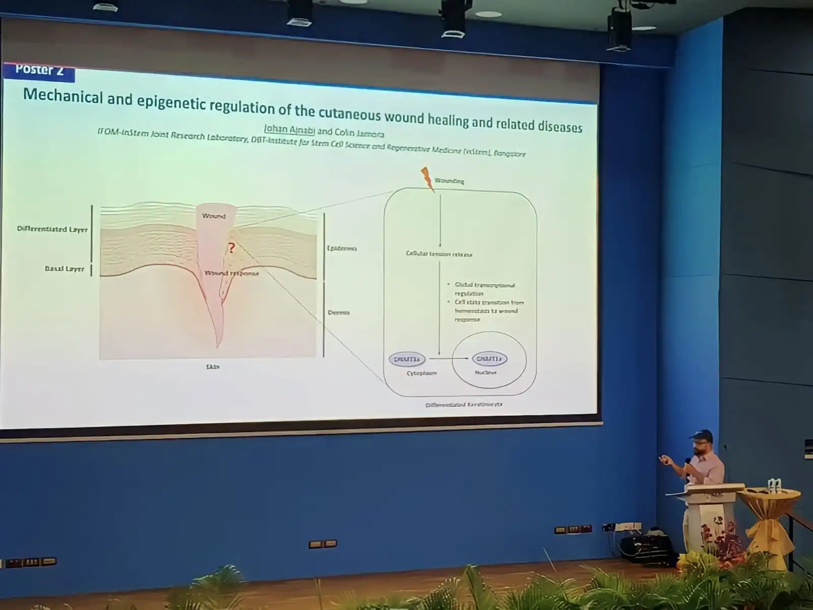
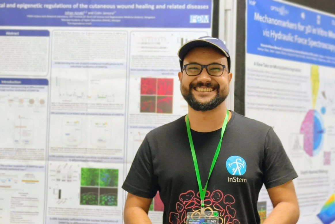
MBI Conference 2023: Mechanobiology in Health and Disease
National University of Singapore (NUS), Singapore · September 2023

Poster Presentation — Epigenetic and mechanical regulation of the cutaneous wound healing response
Additional presentations

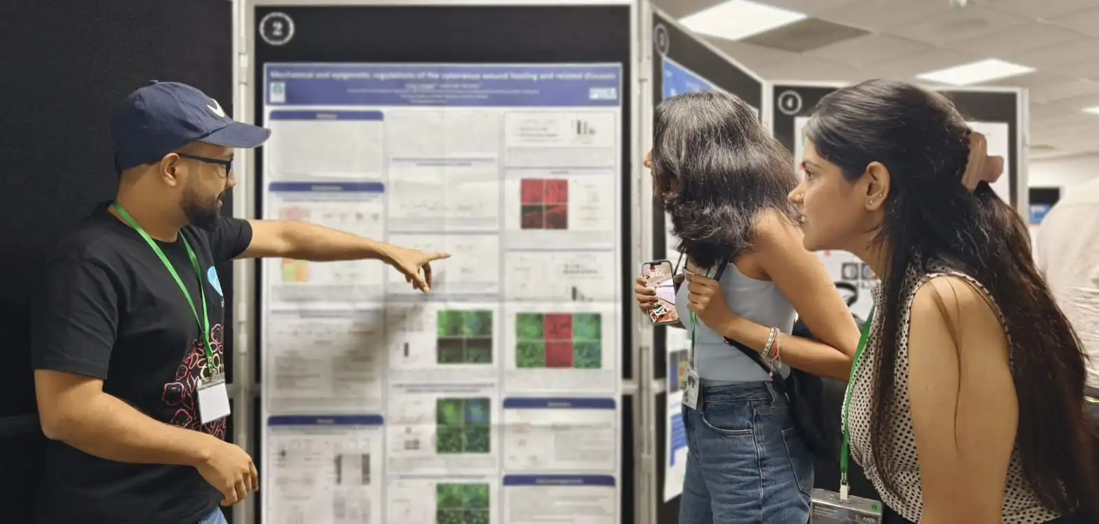
  • Mechanobiology in Translational Research: Bioforces to Bedside
    Department of Bioengineering, Indian Institute of Science (IISc), Bengaluru, India · October 2025
    Poster Presentation — Mechanoregulation of DNMT3a in the cutaneous wound healing response
    Conference details
  • 10th International Conference of Laboratory Animal Scientists' Association (LASA)
    Hyderabad, India · June 2022
    Poster Presentation — Understanding the role of DNMT3A in the cutaneous wound healing response using a mouse model
  • National Agricultural Science Congress
    Delhi, India · February 2019
    Poster Presentation — Identification and characterization of stress responsive PM19 promoter from wheat

↑ Top